

9 min read · 8 November 2026
Heart Disease in Dogs and Cats: Signs, Susceptible Breeds, and What an Echocardiogram Reveals
Cardiac disease is one of the most underdiagnosed conditions in pets — partly because the early signs are easy to miss, and partly because many owners don't know their breed is at risk. Here's what I see at the clinic, and what an echocardiogram can show long before a pet is visibly unwell.
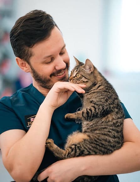
DVM · GP Cert in Ultrasound

More Common Than You Think
One in ten dogs will develop some form of heart disease during their lifetime. In Cavalier King Charles Spaniels, that number climbs to virtually 100% by age ten. In cats, hypertrophic cardiomyopathy is the most common cardiac disease overall — and it can strike apparently healthy animals at any age, sometimes with no warning signs at all until the animal is in crisis.
As a vet with a postgraduate certificate in ultrasound, I perform echocardiograms regularly at Mike's Vet. What consistently surprises owners is how much we can find — and how much can be managed — when cardiac disease is caught early. This article covers what to watch for, which breeds carry the highest risk, how age factors in, and what an echocardiogram actually tells us.
The most important message: by the time heart failure is obvious from the outside — laboured breathing, exercise intolerance, collapse — significant structural damage has often already occurred. Diagnosis before that point gives us far more treatment options and far more time.
Signs to Watch For
Early signs are often subtle and easy to dismiss. A cough first thing in the morning. Slightly reduced tolerance on walks. A pet that seems a little quieter than usual. These can go unnoticed — or be attributed to 'just getting older' — for months.
More specific signs to take seriously: a persistent cough (especially at night or after lying down), faster or harder breathing at rest, reduced stamina, weight loss without a clear cause, abdominal distension (fluid accumulation), fainting or brief episodes of collapse, weakness in the hindquarters.
In cats, the picture is often even less obvious. Cats hide illness well. Some cats present in acute congestive heart failure — literally struggling to breathe — with no prior warning the owner could have detected. This is one reason why breed-based proactive screening matters so much in cats.
One of the most reliable early findings is a heart murmur detected on a routine physical examination. If your vet hears a murmur at a check-up, the recommendation to investigate further is not over-caution — it is early intervention at exactly the right time.
Dog Breeds Most at Risk
Cavalier King Charles Spaniel: the highest-risk dog breed by some margin. Myxomatous mitral valve disease (MMVD) affects virtually all Cavaliers to some degree by age ten [1]. ACVIM consensus guidelines now recommend annual cardiac auscultation from age one, with echocardiography when a murmur is detected. Many cardiologists recommend a baseline echo at three to four years even before a murmur develops, so there is a reference point when disease progresses.
Boxer: prone to arrhythmogenic right ventricular cardiomyopathy (ARVC), also known as Boxer cardiomyopathy [4]. This causes ventricular arrhythmias that can lead to sudden collapse or death — sometimes as the first sign. A Boxer showing episodes of weakness, syncope, or unusual behaviour during exercise warrants prompt evaluation. Holter monitoring over 24 hours is the gold standard for arrhythmia assessment in this breed.
Dobermann Pinscher: dilated cardiomyopathy (DCM) is so prevalent that screening is now recommended even in apparently healthy Dobermanns [2]. DCM in this breed can progress silently for months or years before clinical signs appear. By the time an owner notices anything wrong, the heart may already be significantly enlarged. Echo combined with Holter monitoring is the best screening approach.
Great Dane and other giant breeds: also at significant DCM risk. The sheer size of the heart in giant breeds means that when function declines, it declines fast. Baseline echocardiography from around five to six years of age is advisable in Great Danes, Irish Wolfhounds, and Newfoundlands.
Cocker Spaniel: English and American Cockers share the Cavalier's predisposition to mitral valve disease, though typically with a later age of onset. German Shepherd: DCM and subaortic stenosis (SAS) both occur at higher rates; SAS in particular can cause sudden death in young dogs if severe and undetected.
Other breeds worth noting: Labrador Retriever, Portuguese Water Dog, and Dachshund (mitral valve disease). Smaller crossbreeds with Cavalier heritage — common in Dubai — also carry elevated MMVD risk.
Cat Breeds Most at Risk
Maine Coon: hypertrophic cardiomyopathy (HCM) is the most common cardiac disease in cats overall, and Maine Coons carry a specific genetic mutation (MYBPC3-A31P) that dramatically increases their risk [6]. Genetic testing is available and recommended. Even Maine Coons that test negative for this mutation can still develop HCM through other mechanisms, so negative genetic test results do not eliminate the need for periodic echocardiographic screening.
Ragdoll: another breed with a known HCM-associated mutation (MYBPC3-R820W) [5]. Screening echocardiography from two to three years of age is now considered best practice. Many responsible breeders screen their breeding stock — if you have a Ragdoll and don't know whether the parents were screened, it is worth arranging your own baseline.
British Shorthair: HCM prevalence studies have consistently found British Shorthairs to be over-represented. The breed is popular in Dubai and the UAE generally, and many owners here are unaware of this risk. A routine check-up is not sufficient to rule out early HCM — only an echocardiogram can assess structural changes in the myocardium.
Persian and Exotic Shorthair: moderate to high HCM risk. The brachycephalic conformation in these breeds can mask respiratory symptoms of cardiac disease, making careful echo assessment important — a Persian breathing harder than usual may have cardiac disease, respiratory disease, or both.
Sphynx: HCM and restrictive cardiomyopathy (RCM) both occur at elevated rates in Sphynx compared to the general cat population. Mixed-breed cats: not immune. HCM occurs across all feline breeds and in domestic shorthairs [3]. Any cat with a murmur, respiratory signs, or a breed predisposition warrants cardiac evaluation regardless of pedigree.
Age Matters: When Does Cardiac Disease Typically Develop?
In dogs, most acquired cardiac disease presents in middle age or later. Cavalier King Charles Spaniels typically develop their first murmur between three and five years, with disease progression through the later years. DCM in Dobermanns most commonly manifests between six and ten years. Giant breeds like Great Danes may show cardiac changes from five to six years onward.
Congenital defects — pulmonic stenosis, aortic stenosis, patent ductus arteriosus (PDA), ventricular septal defects — are present from birth and are typically detected during a puppy's first examination. If a murmur is heard in a puppy under six months, it is usually investigated to determine whether it is a congenital defect or an innocent flow murmur that will resolve on its own.
In cats, HCM is less age-predictable. It can occur in cats as young as one to two years, particularly in Maine Coons and Ragdolls. In mixed-breed cats it most commonly presents between six and twelve years. What makes feline cardiac disease particularly challenging is that many cats carry significant structural abnormalities for years without detectable clinical signs — then present in acute respiratory distress.
The practical implication for at-risk breeds: establishing a baseline echocardiogram before the typical age of disease onset gives you a reference point for comparison as your pet ages. For Cavaliers, I recommend this by age three to four. For Maine Coons and Ragdolls, two to three years. For Dobermanns, annually from age five. For other at-risk breeds, discuss timing with your vet based on breed-specific guidance.
What an Echocardiogram Actually Shows
An echocardiogram is an ultrasound of the heart. It allows us to look at the heart's structure and function in real time — watching it beat, measuring chamber sizes, assessing valve movement, and mapping blood flow with Doppler. It is non-invasive, requires no anaesthesia in most cases, and gives us information no physical examination or chest X-ray can provide.
At Mike's Vet, we use the Mindray Vetus9 — one of the most advanced veterinary ultrasound platforms available. Its Zone Sonography Technology (ZST+) delivers exceptional image clarity even in animals with deep chests, and its Tissue Doppler Imaging (TDI) capability lets us detect subtle changes in myocardial function that wouldn't be visible on a standard echo. The dedicated cardiology suite includes pulsed and colour-flow Doppler, continuous-wave Doppler, and automated ejection fraction (Auto EF) measurement.
Specifically, an echocardiogram allows us to assess: the size of each cardiac chamber and whether they are within normal limits; whether chamber walls are thickened as in HCM or thinned and dilated as in DCM; how well the heart muscle is contracting and relaxing; the presence and severity of valve abnormalities; whether blood is regurgitating through a leaking valve and how significant the leak is; the presence of pericardial effusion (fluid around the heart); and signs of elevated filling pressures that indicate impending or early heart failure.
For a Cavalier with a known murmur, the echo tells us which ACVIM stage they are in — which directly determines whether and when to start medication. For a Ragdoll with no symptoms at all, it may reveal significant left atrial enlargement that changes management entirely. For a dog presenting with a cough, it distinguishes cardiac from respiratory causes — a distinction that determines the entire treatment approach.
Most pets tolerate echocardiography well without sedation. The scan takes around 20 to 40 minutes. We clip a small area of fur on the chest, usually just below the elbow joint, to allow the probe to make good contact with the skin. We provide a written report with measurements, images, and — where disease is found — a staged management plan.
Treatment and What It Can Achieve
The most significant advance in small animal cardiology over the past decade has been the evidence from the EPIC trial, which showed that starting pimobendan in dogs with pre-clinical MMVD — specifically those with cardiac enlargement but before symptoms appear — significantly delays the onset of heart failure [1]. This changed the standard of care for Cavaliers and other dogs with mitral valve disease. We now actively look for the echocardiographic thresholds that determine whether a dog qualifies for this intervention.
For cats with HCM, management depends on the stage and presentation. Cats in congestive heart failure require diuretics; some with elevated risk of aortic thromboembolism receive antiplatelet therapy; additional agents may be used in specific cases [3]. The honest reality is that feline HCM management remains more challenging than canine MMVD — but early detection consistently improves outcomes by giving us more time and more options before a crisis develops.
For DCM, management addresses both the myocardial dysfunction and any arrhythmias present. Holter monitoring gives us the full picture of arrhythmia burden over 24 hours — essential for Dobermanns and Boxers where arrhythmia management is a central part of care.
Lifestyle adjustments complement medication in all cardiac cases: restricting intense exercise in certain conditions, managing weight, reducing dietary sodium where appropriate. The goal is always to extend the period before heart failure, reduce symptoms when they develop, and maintain quality of life. With early detection and appropriate management, many cardiac patients live comfortably for years after diagnosis.
Cardiac Assessment at Mike's Vet
If you have a Cavalier King Charles Spaniel, Maine Coon, Ragdoll, Dobermann, Boxer, or British Shorthair — or any breed mentioned in this article — I would encourage you not to wait for symptoms. Book a cardiac check and get a baseline.
Cardiac evaluation at Mike's Vet begins with a thorough auscultation. If we detect a murmur, or if you want a proactive baseline for an at-risk breed, the next step is an echocardiogram on the same day in most cases. We use our Mindray Vetus9 platform, provide a written report with measurements and images, and walk you through what we found and what it means.
Pre-anaesthetic cardiac screening is also advisable before surgery in any at-risk breed or any animal over eight years — we routinely incorporate cardiac assessment into our pre-operative workup for older patients. For Dobermanns and Boxers where arrhythmia evaluation is part of the picture, we can arrange Holter monitoring.
Cardiac workups are available at our Hessa Street clinic, 7 days a week. Book via WhatsApp at +971 4 283 7744 or online at the link below.
Book an appointment at Mike's Vet
Open 7 days · 9 AM – 9:30 PM · Hessa Street, Dubai
Related: X-Ray & Ultrasound Dubai
Learn more →References
- 1.Keene BW, Atkins CE, Bonagura JD, et al. ACVIM consensus guidelines for the diagnosis and treatment of myxomatous mitral valve disease in dogs. J Vet Intern Med. 2019;33(3):1127–1140.
- 2.Keene BW, Atkins CE, Bonagura JD, et al. ACVIM consensus guidelines for the diagnosis and treatment of dilated cardiomyopathy in dogs. J Vet Intern Med. 2019;33(3):1141–1163.
- 3.Luis Fuentes V, Abbott J, Chetboul V, et al. ACVIM consensus statement guidelines for the classification, diagnosis, and management of cardiomyopathies in cats. J Vet Intern Med. 2020;34(3):1062–1077.
- 4.Meurs KM, Spier AW, Miller MW, Lehmkuhl LB, Towbin JA. Familial ventricular arrhythmias in Boxers. J Vet Intern Med. 2001;15(5):437–439.
- 5.Meurs KM, Norgard MM, Ederer MM, Hendrix SM, Kittleson MD. A substitution mutation in the myosin binding protein C gene in Ragdoll hypertrophic cardiomyopathy. Genomics. 2007;90(2):261–264.
- 6.Meurs KM, Sanchez X, David RM, et al. A cardiac myosin binding protein C mutation in the Maine Coon cat with familial hypertrophic cardiomyopathy. Hum Mol Genet. 2005;14(23):3587–3593.
Frequently Asked Questions
How do I know if my dog or cat has a heart problem?
Early signs are often subtle: a persistent cough (especially at night), reduced exercise tolerance, faster breathing at rest, or weight loss without an obvious cause. In cats, signs can be even less obvious — some cats present in acute respiratory distress with no prior warning. If your vet hears a heart murmur on a routine examination, that is the most reliable early finding. For at-risk breeds, proactive echocardiographic screening is recommended even in apparently healthy animals.
What is HCM in cats and which breeds are affected?
Hypertrophic cardiomyopathy (HCM) is the most common cardiac disease in cats. It causes the walls of the heart muscle to thicken, reducing the heart's ability to fill and pump properly. Maine Coons and Ragdolls carry known genetic mutations that increase their risk significantly. British Shorthairs, Persians, and Sphynx are also over-represented. It can occur in mixed-breed cats too, particularly in middle age and older. Early diagnosis with echocardiography allows for monitoring and timely intervention.
My vet heard a heart murmur in my dog — what happens next?
A murmur is a sound caused by turbulent blood flow through the heart. It is graded on a scale of 1 to 6. A low-grade murmur in an older Cavalier may represent early mitral valve disease; the same murmur in a young puppy may be an innocent flow murmur that resolves on its own. The next step is usually an echocardiogram to assess the underlying cause, the severity of any valve abnormality, whether the heart chambers are enlarged, and what ACVIM stage the disease is at. That staging determines whether and when medication is appropriate.
How much does a pet echocardiogram cost in Dubai?
At Mike's Vet, a full abdominal ultrasound is AED 886 and a chest ultrasound is AED 561. Cardiac echocardiography pricing is discussed at the time of booking depending on the scope of assessment needed. Call or WhatsApp +971 4 283 7744 for a quote. We provide a written report with all measurements and images.
Is echocardiography safe for my pet? Do they need to be sedated?
Echocardiography is completely safe — it uses the same ultrasound technology as human cardiac imaging. The vast majority of dogs and cats tolerate the procedure well without any sedation. We clip a small area of fur on the chest to allow the probe to make good skin contact. The scan takes 20 to 40 minutes depending on what we are assessing.
Can heart disease in pets be cured?
Most cardiac diseases in pets are managed rather than cured. Mitral valve disease progresses over time, but medications like pimobendan can significantly delay the onset of heart failure when started at the right stage. HCM in cats is managed with medications that reduce symptoms and risk of complications. Congenital defects like patent ductus arteriosus (PDA) can often be corrected surgically. The goal of management is always to extend comfortable, good-quality life — many cardiac patients do very well for years after diagnosis.